


Summary list for 6.2 Blood system
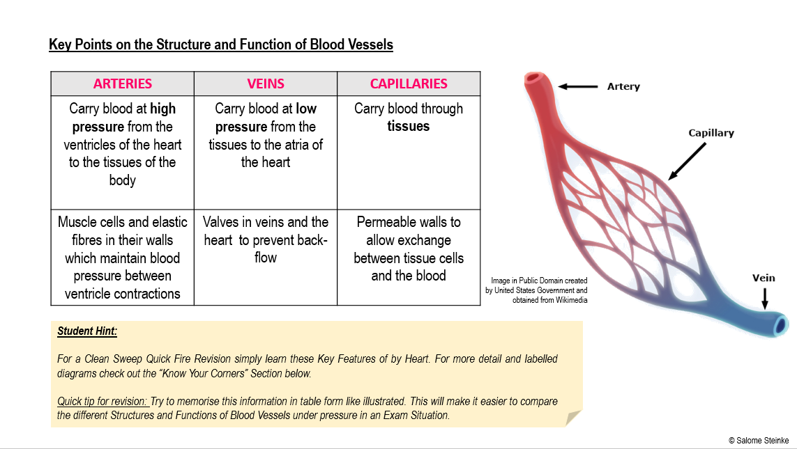
- Arteries carry blood at high pressure from the ventricles of the heart to the tissues of the body.
- Arteries have muscle cells and elastic fibres in their walls.which maintain blood pressure between ventricle contractions.
- Capillaries carry blood through tissues and have permeable walls to allow exchange between tissue cells and the blood.
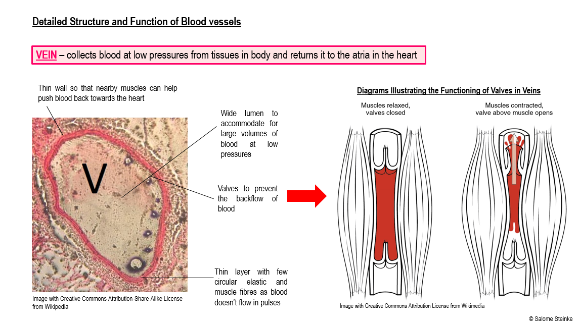
- Veins carry blood at low pressure from the tissues to the atria of the heart.
- Valves in veins and the heart prevent back-flow.
- There is a separate circulation for the lungs.
- The sinoatrial node (SA node) initiates heart beat
- The SA node is made of specialised muscle cells in the right atrium which act as a pacemaker.
- The sinoatrial node sends out an electrical signal which is propagated through the walls of the atria and ventricles.
- The heart rate is controlled by impulses brought to the heart through two nerves from the medulla of the brain.
- Epinephrine (adrenalin) increases the heart rate.
Skills
- Outline your knowledge of William Harvey’s discovery of the circulation of the blood with the heart acting as the pump.
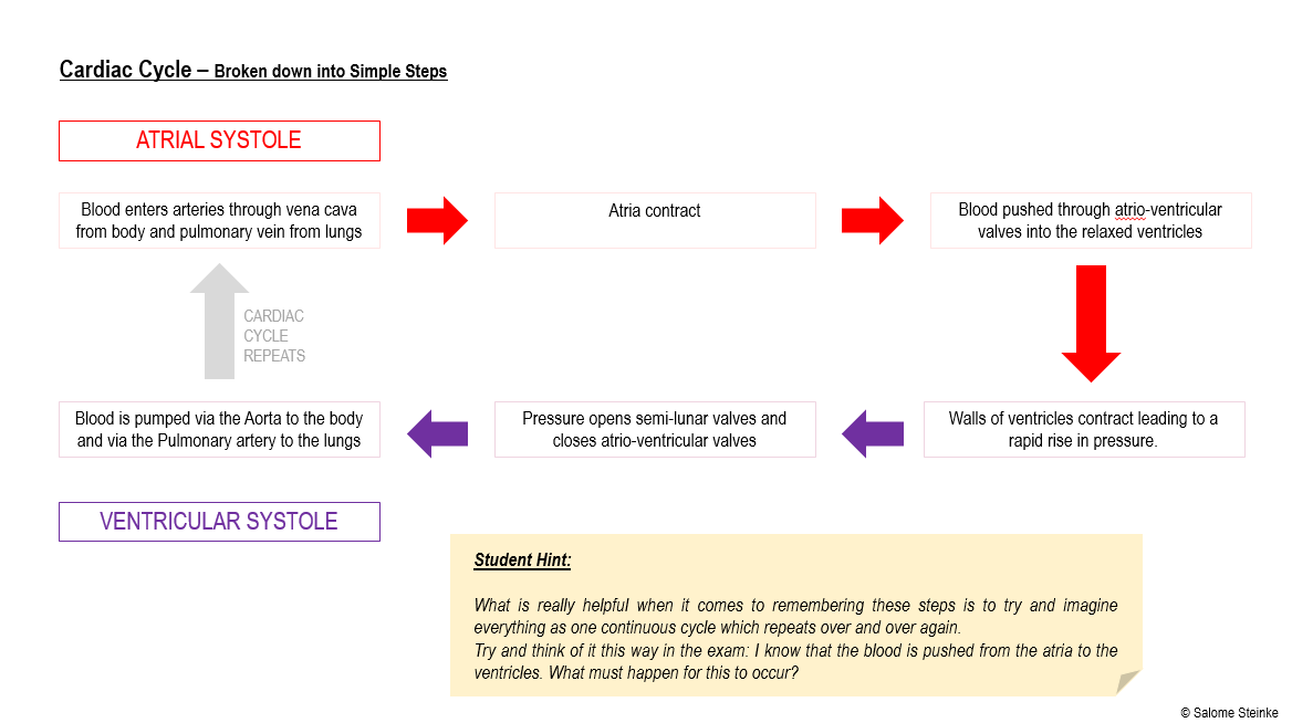
- Interpret pressure changes in the left atrium, left ventricle and aorta during the cardiac cycle.
- Link causes and consequences of occlusion of the coronary arteries.
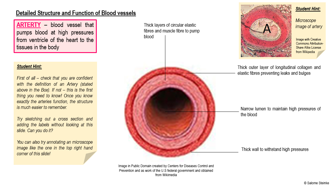
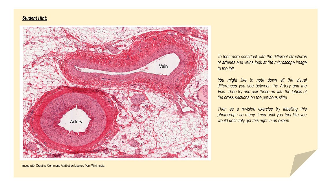
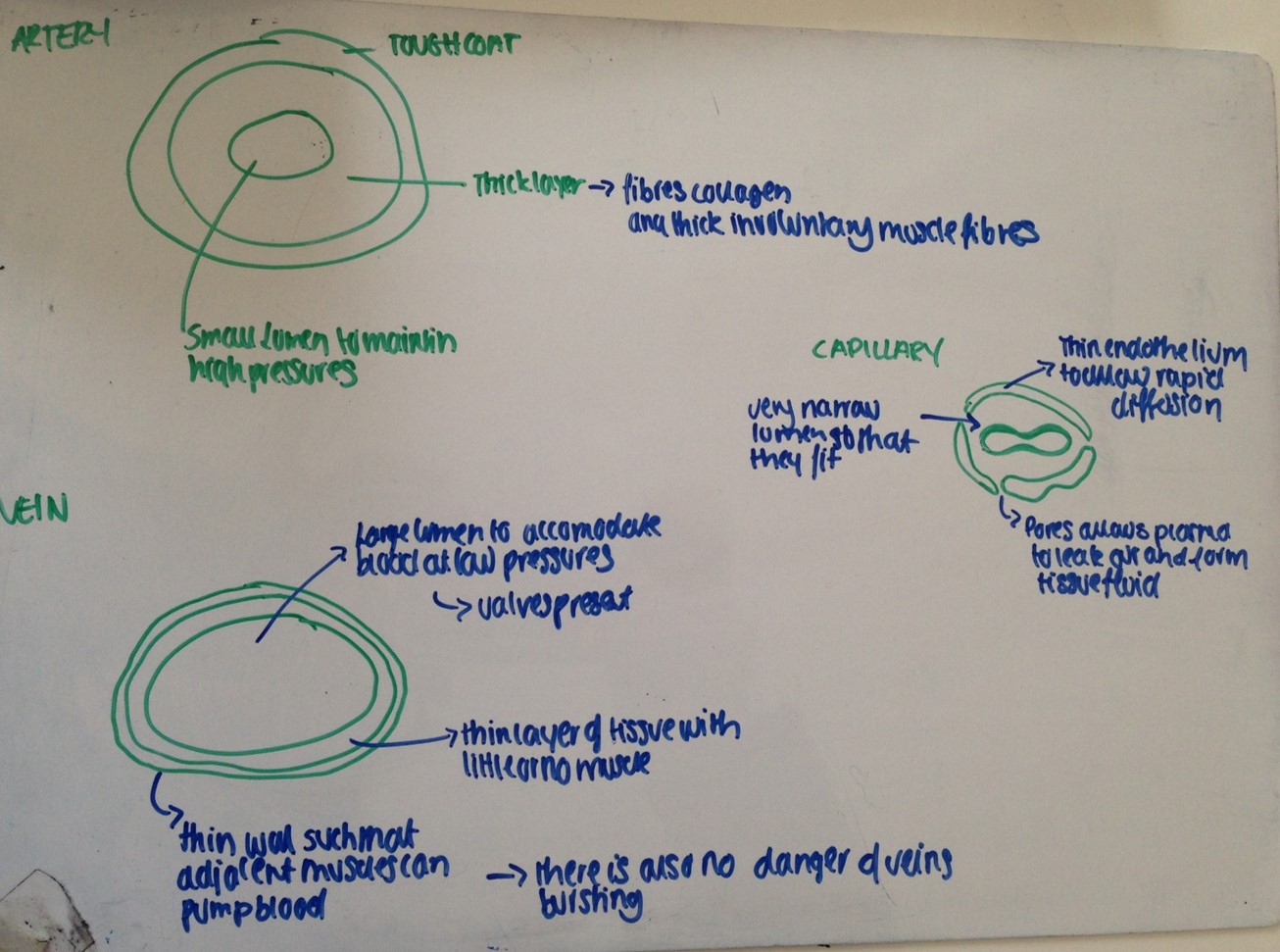
- Identify arteries, capillaries or veins from the structure of their walls.
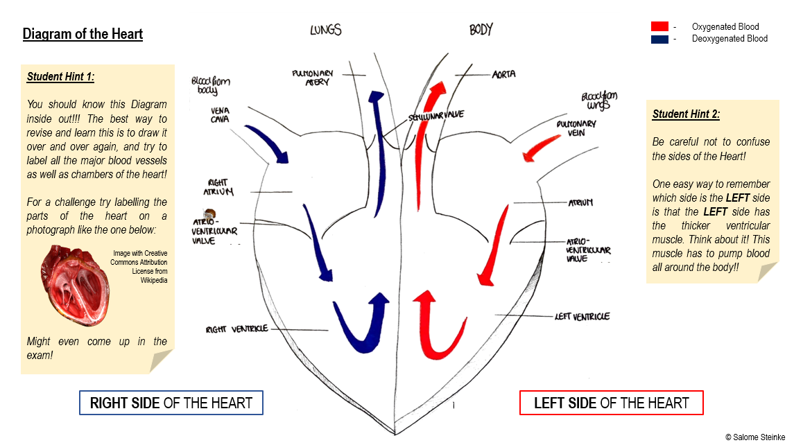
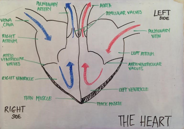
- Recognise the four chambers and valves of the heart and the blood vessels connected to it in a dissected heart or in diagrams of heart structure.
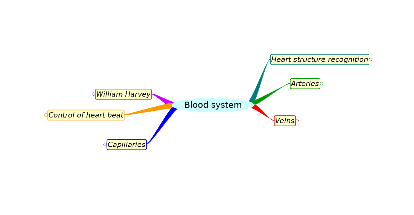
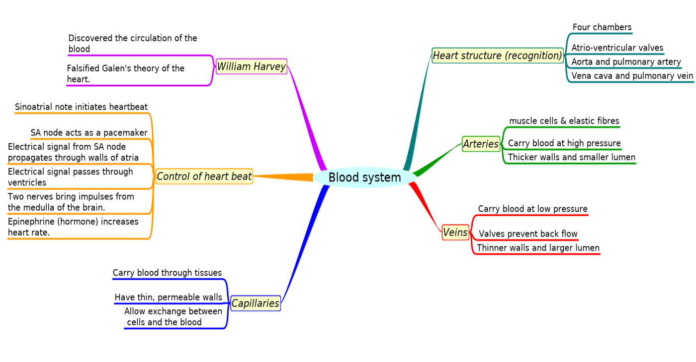
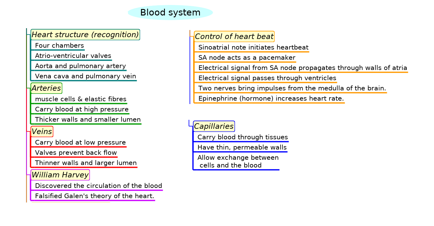
Mindmaps
This diagram summaries the main sections of topic 6.2.
Test if you can draw something like these concept maps from memory.


Revision Exercise - Whiteboards
Do you really need to revise but you don't feel like copying out all your notes?
Want something quicker and more effective? All you need is a small whiteboard and pen.
Instead of laboriously writing out all of your notes (particularly diagrams!) try sketching. This technique is not only faster than writing on paper but also allows you to easily make changes, rub things out or add things you may have forgotten.
Doesn't have to be that neat either! But don't forget to take a picture of all of your whiteboard "pages".
They can be useful revision notes to come back to later! Check out two examples below..
Exam style question about pressure changes in the heart
Explaining the flow of blood through the heart and the opening and closing of valves is an important skill in this topic.
Answer the question below, on a piece of paper, then check your answer against the model answer below.
The image shows a diagram of the heart in one stage of the cardiac cycle.
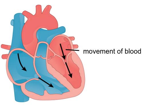
Outline how the contraction and relaxation of muscles in the heart causes different pressures of blood in different chambers leading to blood flow and valve movements in this heart [4]
....................................................................................... ............................................................................
....................................................................................... ............................................................................
....................................................................................... ............................................................................
....................................................................................... ............................................................................
Click the + icon to see a model answer.
This question tests an understanding of the structure and function of arteries and veins.
Answer the question below on a piece of paper, then check your answer with the model answer.
The diagram shows a cross section of an artery and a vein.
Label the image with an X to identify which vessel is an artery giving one reason. (2 marks)
Explain how the structure of the artery is related to its function in the circulatory system. (4 marks)
Examiner hints:
Identify- give an answer from several possibilities
Explain includes reasons
Click the + to see a model answer.
This is a self marking quiz containing questions covering the topic outlined above.
Try the questions to check your understanding.
START QUIZ!
T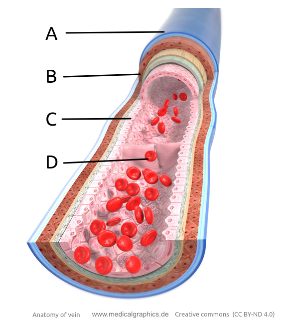 he image shows a model of a vein.
The layers are shown as well as blood cells and a valve.
Which label points to the muscle layer of the vein?
Layer B is the muscle layer. C is the endothelium, and D is the valve.
An artery and a vein can be seen in the image.
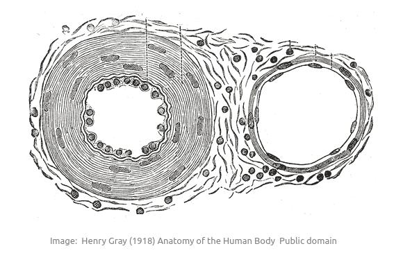
Which row in the table below correctly describes
differences between arteries and veins?
| Artery | Vein | |||
| Muscle layer | Lumen | Muscle layer | Lumen | |
| A | large thickness | small | large thickness | small |
| B | medium thickness | medium | small thickness | very small |
| C | very large thickness | medium | medium thickness | large |
| D | medium thickness | small | very large thickness | medium |
The muscle layer is the thickest layer in both artery and vein, but arteries have a thicker muscle layer than veins, and also a thicker outer layer rich in elastic fibres.
The order of size of the terms used in the table, starting with the biggest is; very large, large, medium, small, very small
The control of heart rate involves several body systems.
Which of the following can increase heart rate?
I: Nerve impulses from the medulla of the brain
II: Depolarization of the heart atria muscle cells
III: Release of the hormone leptin
IV: Release of the hormone epinephrine
The SA node is the pacemaker of the heart, it's activity can be increased using nerves impulses from the brain, and by the release of the hormone epinephrine (adrenalin)
he heart is said to be myogenic. Which part of the heart is responsible for creating the heart's natural electrical rhythm?
The sinoatrial node (SA node) sends out an electrical signal in a regular rhythm which stimulates the contraction of the atria then the ventricles.
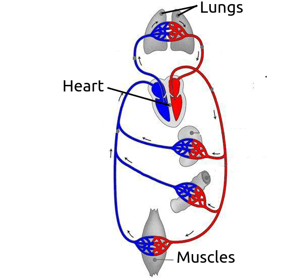 The image shows a simplified diagram of the human circulatory system.
It shows the general layout of circulation to and from the heart.
What do the blue and red vessels represent?
The human circulation is a double circulation. The blood is pumped twice.
Blue represents deoxygenated blood and red represents deoxygenated blood.
How is a heart beat initiated in the heart?
The SA node is made from a group of specialised muscle cells. This is found in the right atrium wall.
Cardiac muscle is special in that it can conduct electrical impulses as well as contracting.
Why do arteries have thick muscular walls and elastic fibres?
The arteries receive blood from the heart. When the ventricles contract the blood pressure rises and becomes pressurised, stretching the arteries which need strong thick walls to resist this pressure.
Which of the following statements best describes the structure and function of veins?
The role of veins is to return the blood to the heart. The pressure is quite low, and valves prevent the back flow. The lumen is large and the walls are quite thin because there is no need to withstand high pressure.
Drag and drop activities
Test your ability to construct biological explanations using the drag and drop questions below.
The heart beat is controlled by the pacemaker but the rate can be increased or decreased depending upon the needs of the body.
Drag and drop the correct word or phrase into the gap to describe the control of the heart rate.
increase increased pacemaker normal heart rate slow cardiovascular centre accelerans fear ventricles sinoatrial node contraction involuntarily contraction right voluntarily left atrium epinephrine
Heart muscle is myogenic, it contracts without stimulation. The heart has a in the atrium which generates the at approximately 72 beats per minute without nervous or hormonal control.
The pacemaker, the , initiates the heart beat and of the heart muscle starting from the right atrium, passing through the and then to the causing of the heart muscle in the correct sequence.
The rate of the heartbeat can be or decreased by the of the medulla oblongata in the hind brain utilizing the vagus nerve to the heart (rest) and the nerve to the heart rate (exercise).
The hormone, , which is released in a stress or situation to stimulate the “fight or flight” response also increases the heart rate.
The heart beat is controlled by the sinoatrial node pacemaker but the rate can be increased or decreased by nerves and hormones to suit the needs of the body.
This is a little bit of fun. You can choose your favourite game.
If you can't see the content below please click this direct link to Blood system card matching game.
How much of Blood system 6.2 have you understood?
 Twitter
  Facebook
  LinkedIn