Question 20N.3.SL.TZ0.b
| Date | November 2020 | Marks available | [Maximum mark: 2] | Reference code | 20N.3.SL.TZ0.b |
| Level | SL | Paper | 3 | Time zone | TZ0 |
| Command term | Explain | Question number | b | Adapted from | N/A |
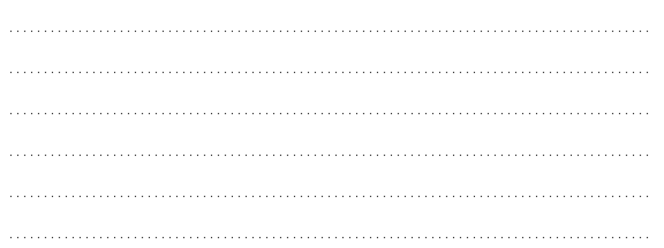
The micrograph shows a section through part of the liver. The diagram illustrates the details of what is found within the marked area.

[micrograph] Micrograph of liver. https://undergraduate.vetmed.wsu.edu/courses/vph-308/histology/lab-1-histologycells-
and-organelles/liver-slide-wsu_2_052 courtesy of Patrick D. Wilson, MS, DVM, Clinical Associate Professor,
Veterinary Integrative Biosciences, College of Veterinary Medicine & Biosciences, Texas A&M University.
[diagram] Reprinted by permission from Springer Nature, Nature Reviews Immunology, “Aberrant homing of mucosal
T cells and extra-intestinal manifestations of inflammatory bowel disease” by Adams and Eksteen ©2006. https://www.nature.com/nri/.
Explain the function of hepatocytes in protein metabolism.
[2]
a. they produce/secrete plasma proteins;
b. (the plasma proteins) are modified/secreted by the Golgi apparatus;
c. protein/globin is broken down into amino acids;
The function of the hepatocytes in protein metabolism was challenging for most candidates despite this being taken directly from an assessment statement.